Eye Health

Learn about the eye’s anatomy and common eye diseases — including causes, symptoms, and options for prevention and treatment.
 

Good eye health and eye care are important to protect your sight, prevent common eye diseases and treat eye problems early.

The best way to maintain your best vision is by living an overall healthy lifestyle. This includes protecting your eyes from the sun’s ultraviolet radiation, not smoking, eating a healthy diet, getting plenty of physical activity, and having regular, comprehensive dilated eye exams. Check out our Comprehensive Dilated Eye Exam page to learn more about this sight-saving exam.

Here, take a tour of the eye to learn how the eye works. Find out about common eye conditions, as well as causes, risk factors, symptoms, detection and treatment options, and current research.

Tour of the Eye

How Does the Eye Work?

The eye is an organ with several parts. These parts work together to perform the eye’s primary function: to detect light. This section features information about these parts and how they can impact vision.


Eyelids

The eyelids protect the eye. Glands in the eyelid secrete oil, water, and other substances, which are spread in an even layer across the surface of the eye, forming the “tear film,” when the eyelids blink. When the glands are unhealthy, the tear film is missing components, and the eye may feel dry and scratchy or have excess tearing. Treatments for this condition include using warm compresses and lubricant eye drops (artificial tears). It is best to use eye drops that are not labeled as “taking the red out.”

Illustration of parts of the eye, indicating position of the upper, lower, and third eyelids


Sclera

The white, outer part of the eyeball is called the sclera. It protects the eye.

Illustration of parts of the eye, indicating position of the sclera.


Cornea

The cornea is the clear outer part at the front of the eye. It does most of the eye’s focusing. The cornea’s inner layer of cells continuously pumps water out of the cornea. If those cells are damaged, the cornea will become swollen and cloudy, and a corneal transplant or other surgery is needed to restore clear vision. Corneal transplants usually are successful because the cornea has no blood vessels. There is a constant need for organ donors to provide corneas.

Illustration of parts of the eye, indicating position of the cornea


Iris

The iris is the ring-shaped, colored part of the eye behind the cornea. It regulates how much light enters the eye.

Illustration of parts of the eye, indicating position of the iris


Pupil

The pupil is the opening at the center of the iris. The iris’ muscles change the iris’ width, which changes the pupil’s size. That is how the iris controls how much light can enter the eye.

Illustration of parts of the eye, indicating position of the pupil


Lens

The lens helps to focus light onto the retina at the back of the eye. A cloudy lens is called a “cataract.” Cataract surgery can be done when a person notices that his or her vision is inadequate to do whatever he or she likes to or must do. The surgeon makes a tiny incision and inserts a long, thin instrument that breaks up the cloudy lens with ultrasound waves and suctions the pieces out of the eye. A clear, plastic lens is then placed in the eye. The incision is so small that stitches usually are not needed.

Illustration of parts of the eye, indicating position of the lens


Vitreous Gel

The vitreous is a clear gel that fills the inside of the eye behind the lens and helps maintain the round shape of the eye.

Illustration of parts of the eye, indicating position of the vitreous gel


Retina

The retina is a light-sensitive tissue that lines the back of the eye. It converts light into electrical impulses that are sent to the brain through the optic nerve. Many eye diseases affect the retina and thus cause poor vision. Diabetes can cause the growth of abnormal blood vessels on the retina’s surface. These can leak and bleed, and over time they may contract and detach the retina from the back of the eye. Diabetes also can cause swelling of the retina. These problems are treated most quickly and most effectively when caught early. So, everyone with diabetes should have a comprehensive dilated eye exam once a year. The best way to prevent vision problems from diabetes is to keep the blood sugar at a healthy level.

Illustration of parts of the eye, indicating position of the retina


Macula

The macula is a small area of the retina at the center of the back of the eye. It provides central vision. At the center of the macula is the fovea, which allows you to see details sharply. Age-related macular degeneration (AMD) affects the macula. There are two forms: dry and wet. In the wet form, abnormal blood vessels grow beneath the retina. They can leak blood and other substances, and resultant scarring causes loss of central vision. In the dry form, the macula thins and deposits of fat combined with protein (called “drusen”) occur beneath the retina. Dry AMD can become wet AMD, but not vice versa. Both forms of AMD can cause loss of central vision, but wet AMD causes more rapid loss of vision.

Illustration of parts of the eye, indicating position of the macula


Optic Nerve

The optic nerve is a cable of nerve fibers that connects your eye to your brain. The nerve communicates input for the retina to the brain, which tells you what you are seeing. Glaucoma is a group of eye diseases that damage the optic nerve. This damage is irreversible and progresses unless the glaucoma is treated. Treatment does not restore vision lost from glaucoma. Rather, it prevents further damage from occurring, so catching glaucoma early and maintaining treatment for it is important.

Illustration of parts of the eye, indicating position of the optic nerve

Diseases and Conditions

Learn about different types of diseases that commonly affect the eye. Gain a better understanding of signs to look for, factors that can put you at risk, and ways to prevent and treat these conditions.


Amblyopia icon

Amblyopia and Strabismus

After reading the information below, click here to learn more about POB’s programs for children.

Amblyopia, commonly known as “lazy eye,” affects approximately 2 to 3 out of every 100 children. Vision in one of the eyes is reduced because the eye and the brain are not working together correctly. One of amblyopia’s most common precursors is strabismus — otherwise known as “crossed eyes” or “turned eye” — in which an individual’s eyes do not line up in the same direction when focusing on an object.
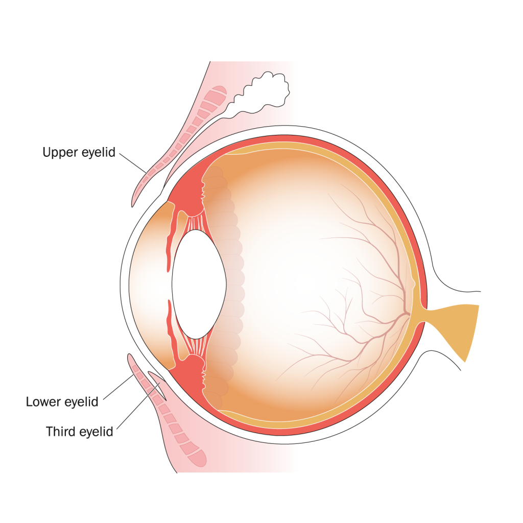
What is Amblyopia?

Amblyopia (“lazy eye”), is decreased vision in one or both eyes due to abnormal vision development in infancy or childhood, causing the brain not to receive normal stimulation from one or both eyes. The eye itself may look normal, but it is not being used normally because the brain is favoring the other eye. The reason it is often called “lazy eye” is because one eye is not being used as much as the other eye.

Without early detection and correction, a child faces a permanent loss of vision in the affected eye.

Treatments are generally most successful when they are done early in life, due to the plasticity of a child’s visual system. At a young age, vision connections from the eyes to the brain are still developing and can, therefore, be corrected. As a child ages, the connections are solidified and cannot be changed. Consequently, all children in the region must be screened by a trained professional using a medical screening model.

Long-term Consequences of Untreated Amblyopia:

  • Permanent vision loss, including loss of depth perception and peripheral vision, may occur in the affected eye if not treated properly.
  • Increased risk of permanent damage to the unaffected eye due to higher usage and exposure.

Causes and Risk Factors:

  • Refractive errors like nearsightedness, farsightedness, and astigmatism. Treatment with prescription eyeglasses or contact lenses can correct these issues.
  • Strabismus (a turned or crossed eye), can cause the affected eye to not work like the straight eye. An eye can drift vertically, or to either side.
  • Children with born congenital cataract are at higher risk for amblyopia. A cataract can cause cloudiness in the lens of an eye, which will cause images to look blurry. This is rarer in babies and children, but still possible.

Symptoms

It can be challenging to spot a child with amblyopia. Depth perception can be an issue. You may also notice your child squinting more, shutting one eye, or tilting their head. Regular (at least annual) vision screenings for young children (ages 1–5) are essential for early detection and treatment.

Treatment

Unless treated in early childhood, amblyopia usually persists into adulthood and will continue to worsen in the affected eye. It is the most common cause of monocular (one- eye) visual impairment among children and young and middle-aged adults. The good news is that early treatment works well and usually prevents long-term vision problems.

If there is an underlying condition, doctors may recommend:

  • Glasses or contacts (for kids with refractive error)
  • Surgery (for kids with cataract)

Then, the doctor will want to “re-train” the brain to use the weaker eye. Like with exercise, the more the eye is used, the stronger it gets. These treatments include:

  • Wearing an eye patch on the stronger eye. This forces your child to use the weaker eye. The length of time child needs to wear the eye patch can vary, and this can be a stressful time for some children. You must be a positive support for your child in helping them comply with the doctor’s instructions.
  • Applying special eye drops in the stronger eye. These eye drops (atropine) temporarily blur a child’s near vision in the stronger eye, forcing the brain to use the weaker eye.

Both of these options can be effective, and treatment length may range from a few weeks to many months. Working with your pediatric ophthalmologist is best to decide which is best for your circumstance.

What is Strabismus?

Strabismus is the medical term used when the two eyes do not look in the same direction at the same time. Also called “crossed eyes” or “turned eye,” the condition occurs in approximately 2 to 4% of the population.

There are three common types of strabismus:

  1. Crossed eyes (Esotropia). A child may be born with this condition, or they may develop it within a few months of birth or around two years of age.
  2. Walleye, or divergent eyes (Exotropia). A child may be born with this condition, or it may develop later.
  3. Vertical strabismus. The eyes are out of alignment vertically.

Long-term Consequences of Untreated Strabismus:

  • Reduction of vision (amblyopia) in the turned eye. Reduced vision may occur in one eye, especially under certain circumstances, such as late treatment.
  • Defective binocular vision. The eyes must be straight for the brain to be able to combine what the two eyes see into a single image. This enables accurate vision and stereopsis (3-D vision), which is used to judge depth.

Causes and Risk Factors:

  • Family history. Most commonly, a tendency to have some type of strabismus is inherited.
  • Farsightedness. Sometimes, the condition is due to the eyes being far-sighted and requiring corrective eyeglasses.
  • Muscle abnormality. The muscles in the eyes may not be working correctly, which can cause strabismus.
  • Another eye problem. Very rarely, strabismus may be secondary to a severe abnormality inside the eye, such as a cataract or tumor.

Symptoms

It can be challenging to spot a child with strabismus. The most common symptom a parent may notice is the misalignment of one or both of their child’s eyes. The child likely does not realize that one or both of their eyes are misaligned. You may notice your child bumping into things more than usual because depth perception can be an issue. You may also see your child squinting more, shutting one eye, or tilting their head. Regular (at least annual) vision screenings for young children (ages 1–5) are essential for early detection and treatment.

Treatment

Treatment aims to restore functional vision in each eye individually, as well as good binocular vision—the earlier the treatment, the better the chances of positive outcomes for the child. If treatment is delayed, vision may not be restored. This permanent vision loss can be prevented with early detection and treatment. Do not delay if your child has strabismus. Seek professional advice from your family doctor.

Treatment usually includes:

  • Patching. Patching the eye that is always straight to bring the vision up to normal in the turned eye.
  • Prescription eyeglasses. Eyeglasses may be used, particularly for eyes that are out of focus.
  • Eye drops. Special eye drops (phospholine iodide) may also help straighten the eyes.
  • Surgery. Surgery on the eye muscles is sometimes necessary.

Mitigating the Effects of Strabismus and Amblyopia

Amblyopia and strabismus can both lead to irreversible vision loss in children. The best way to prevent this is through early detection and treatment of these diseases. The American Academy of Ophthalmology and the American Association for Pediatric Ophthalmology and Strabismus offer age-specific recommendations for childhood eye screening:

  • Newborn: An ophthalmologist, pediatrician, family doctor, or another trained health professional should examine a newborn baby’s eyes.
  • Infant: A second screening for eye health should be done by an ophthalmologist, pediatrician, family doctor, or another trained health professional at a well-child exam between six months and the first birthday.
  • Preschool-Age: Between the ages of 3 and 3½, a child’s vision and eye alignment should be assessed by a pediatrician, family doctor, ophthalmologist, optometrist, or person trained in vision assessment of preschool children.
  • Visual acuity should be tested as soon as the child is old enough to cooperate with an eye exam using an eye chart.
  • School-age: Upon entering school, or whenever a problem is suspected, children’s eyes should be screened for visual acuity and alignment by a pediatrician, family doctor, ophthalmologist, optometrist, orthoptist or person trained in vision assessment of school-aged children, such as a school nurse.

However, if you or your child notices problems with his or her vision, visit an eye doctor immediately.

Events

No event found!


Cataract icon

Cataracts

What is a Cataract?

A cataract is a clouding of the eyeÆs lens that affects vision. Most cataracts are related to aging. Cataracts are incredibly prevalent; by age 80, more than half of all Americans either have a cataract or have had cataract surgery.

Causes

The eye’s lens has proteins in it that will begin breaking down and clumping together as we age. This creates the clouding, which worsens over time. A cataract can occur in either one eye or both eyes. It cannot spread from one eye to the other.

Symptoms

Your eye doctor will check for cataracts during a dilated eye exam. However, you may notice signs of cataracts before an exam. If you experience any of the following, be sure to mention them to your doctor. Symptoms include:

  • Cloudy or blurry vision
  • Fading of colors
  • Poor night vision
  • The appearance of halos around lights or lights appear too bright

Treatment

Cataracts are removed surgically. Surgery may not be recommended in the early stages, but when the cataract begins to affect your quality of life significantly, surgery will be necessary.

Cataract surgery is one of the safest surgeries available. The surgeon makes a tiny incision and inserts a long, thin instrument that breaks up the cloudy lens with ultrasound waves and suctions the pieces out of the eye. A clear plastic lens is then placed in the eye. The incision is so small that stitches are usually not needed. This lens can also be specialized based on your eyeglasses prescription, which can further improve your eyesight.

How can I slow or prevent cataracts?

  • Wear sunglasses with UV protection and a hat outside
  • Quit smoking if you are a smoker
  • Eat a healthy diet including fruits and vegetables
  • Have regular comprehensive dilated eye exams

Other Types of Cataract

While most cataracts are related to aging, there are other types:

  • Secondary cataract. Cataracts that form after surgery for other eye problems, such as glaucoma. Cataracts can also develop in people who have other health problems, such as diabetes or conditions that are treated with prolonged steroid use.
  • Traumatic cataract. Cataracts can develop after an eye injury, sometimes years later.
  • Congenital cataract. Some individuals are born with cataracts or develop them in childhood, often in both eyes. These cataracts may be so small that they do not affect vision. If they do, the lenses may need to be removed.
  • Radiation cataract. Cataracts can develop after radiation exposure.

Events

No event found!


Diabetic retinopathy icon

Diabetic Retinopathy

After reading the information below, click here to learn more about POB’s programs for people with diabetic retinopathy.

What is Diabetic Retinopathy?

Diabetic retinopathy is the most common diabetic eye disease and a leading cause of blindness in American adults. Uncontrolled blood sugar levels in the body can cause damage to blood vessels in the retina — a film in the back part of your eye that detects light and sends signals to the brain via the optic nerve. This damage can cause fluid leakage or bleeding. In addition, damaged blood vessels may lead to the development of abnormal growth of new blood vessels on the surface of the retina.

In the disease’s early stages, a person may not notice changes to his or her vision. But over time, diabetic retinopathy can get worse and cause vision loss. Diabetic retinopathy usually affects both eyes. A comprehensive dilated eye exam can catch the disease in its early stages and get you on the road to treatment to prevent or slow permanent vision loss.

What are the stages of Diabetic Retinopathy?

Diabetic retinopathy has four stages:

  1. Mild Nonproliferative Retinopathy. At this stage, microaneurysms occur. These are small areas of balloon-like swelling in the retina’s tiny blood vessels.
  2. Moderate Nonproliferative Retinopathy. This stage is when blood vessels that nourish the retina are blocked. Hemorrhages also appear in the retina.
  3. Severe Nonproliferative Retinopathy. In this stage, many more blood vessels are blocked, depriving several areas of the retina of their blood supply. The amount of hemorrhage in the retina also increases significantly.
  4. Proliferative Retinopathy. At this advanced stage, new and abnormal blood vessels grow on the retina’s surface. These new blood vessels are fragile and tend to bleed, filling the eye with vision-threatening hemorrhage. They can also turn into scar tissue, which contracts over time and causes the retina to detach, leading to blindness.

Symptoms

Early stages of diabetic retinopathy often don’t show any symptoms. You can only detect the disease in these early stages through a comprehensive dilated eye exam. As the disease progresses, you may find it more difficult to see during some activities like reading or driving, and these issues may come and go. In later stages, you may begin seeing dark, floating spots or “cobwebs” that could stay or clear up over time. It is very important to see your eye doctor anytime you experience visual symptoms. You may need prompt treatment to prevent permanent vision problems.

Risk Factors:

  • Diabetes: Type 1, Type 2
    • The longer you have diabetes, the higher your risk. The risk is especially significant in those with uncontrolled blood sugar levels. Two in five Americans with diabetes have some stage of the disease.
    • Women with diabetes who become pregnant or who develop gestational  diabetes are at higher risk. Get a comprehensive dilated eye exam if this is you.

Treatments:

In the early stages of diabetic retinopathy, there is no treatment except preventive strategies and close monitoring by your eye doctor. Regular comprehensive dilated eye exams will help monitor or catch early signs of the disease.

Other prevention methods:

  • Control your blood sugar levels and manage your diabetes.
  • Maintain a healthy diet and get regular physical activity.
  • Maintain healthy blood pressure and cholesterol levels.

In later stages, there are a few treatment options:

  • Injections. Anti-VEGF drugs, such as those used in age-related macular degeneration, can help reverse or slow the leakage of the blood vessels and growth of abnormal blood vessels.
  • Laser Treatments. Your eye doctor may use lasers to shrink or stop leakage of blood vessels and to control the growth of abnormal blood vessels.
  • Surgery. A vitrectomy can help doctors find and repair sources of bleeding in the retina, clear bleeding from inside the eye, and repair detached retina.

Conditions Resulting from Diabetic Retinopathy:

  • Diabetic Macular Edema (DME). Many individuals with diabetic retinopathy will develop DME over time. Blood vessels in the retina leak fluid, causing swelling in the macula and blurry vision. This is the most common cause of visual loss among people with diabetic retinopathy.
  • Retinal Detachment. Diabetic retinopathy can cause scarring of the retina, which can cause the retina to pull away from the back of the eye.
  • Neovascular Glaucoma. Abnormal blood vessel growth is possible in diabetic retinopathy, which can block the fluid draining system of the eye, causing glaucoma.

Events

No event found!


Dry eye icon

Dry Eye

What is Dry Eye?

Dry eye occurs when the eye does not produce tears properly, or when the tears are not of the correct consistency and evaporate too quickly. This will make your eyes feel uncomfortable and can cause vision problems. Dry eye can make many activities, such as using a computer or reading for an extended period, much more difficult. It can also decrease tolerance for dry environments, such as inside an airplane.

Other names for dry eye include dry eye syndrome, keratoconjunctivitis sicca (KCS), dysfunctional tear syndrome, lacrimal keratoconjunctivitis, evaporative tear deficiency, aqueous tear deficiency, and LASIK-induced neurotrophic epitheliopathy (LNE).

Dry eye is common, affecting millions of Americans each year. There are many things you can do to help alleviate the issue and keep your eyes healthy!

What are the types of dry eye?

Aqueous tear-deficient dry eye is a disorder in which the lacrimal glands fail to produce enough of the watery component of tears to maintain a healthy eye surface.

Evaporative dry eye may result from inflammation of the meibomian glands, also located in the eyelids. These glands make the lipid or oily part of tears that slows evaporation and keeps the tears stable. With this form of dry eye, you may have excessive tears —  but they are not the correct consistency.

Symptoms:

  • Scratchy eyes (like there is something in your eye)
  • Burning or stinging feelings
  • Red eyes
  • Sensitivity to light
  • Blurry vision

Inflammation of the surface of the eye may occur along with dry eye. If left untreated, this condition can lead to pain, ulcers, or scars on the cornea, and some loss of vision. However, permanent loss of vision from dry eye is uncommon.

Risk Factors:

  • Being female
  • Being over age 50
  • Poor diet
  • Autoimmune diseases
  • Wearing contact lenses

Treatment

Treatments for dry eye vary, depending on the underlying reasons and type of dry eye  you have. It is vital to talk with your doctor about your options and communicate with them anything you have tried or want to try.

Certain lifestyle changes can help prevent dry eye:

  • Avoid smoke, wind and air conditioning directly on the eyes.
  • Limit screen time and take eye breaks. Use the 20-20-20 rule: every 20 minutes, look at something 20 feet away for 20 seconds.
  • Use a humidifier in places where you spend most of your day — the office, in front of your computer, etc.
  • Drink more water.
  • Sleep more; 7 – 8 hours a night!

If you already have dry eye, the following treatments are available:

  • Over the counter eye drops (artificial tears) are the most common treatment for mild dry eye. This is not the tears to “get the red out,” which are not helpful for dry eye and may exacerbate symptoms.
  • Prescription medicines are available for severe cases of dry eye.
  • For those with excessive, watery tears, warm compresses can be helpful. These compresses help open the clogged glands that contain the oils your tears need. Many warm compress pads are available online.
  • Tear duct plugs may be necessary if tears drain too quickly. “Punctal plugs” can be inserted by your ophthalmologist.
  • In rare, severe cases, there are surgeries available. Often, this will have to do with your eyelids needing to be corrected.

Events

No event found!


Glaucoma icon

Glaucoma

After reading the information below, click here to learn more about POB’s programs for people with glaucoma.

What is Glaucoma?

Glaucoma is a disease of the optic nerve. The hallmark is a progressive loss of nerve tissue over time. In most forms of glaucoma, the damage occurs very slowly, and many patients do not notice a loss of vision until very late in the disease, when most of the nerve has been lost. Because of this, glaucoma has been called the “sneaky thief of sight.” Glaucoma can be treated by lowering the pressure in the eyes, but damage cannot be reversed. This makes early diagnosis and regular follow-up visits to the eye care specialist critical to maintaining vision in patients at risk for glaucoma.

The best way to prevent permanent vision loss from glaucoma is with regular, comprehensive dilated eye exams — the only way to catch the disease before permanent damage occurs.

What is the optic nerve?

The optic nerve is a bundle of more than 1 million nerve fibers. The optic nerve connects the retina to the brain. The retina is the light-sensitive tissue at the back of the eye. A healthy optic nerve is necessary for good vision.

What are the types of glaucoma, their symptoms and their treatments?

Open-angle glaucoma: The most common type of glaucoma. Fluid in the eye does not drain properly, causing elevated eye pressure for the specific eye, pushing on the optic nerve, causing damage. The only way to catch the disease before vision loss occurs is through regular, comprehensive dilated eye exams.

  • Symptoms: Referred to as the “sneaky thief of sight,” open-angle glaucoma often has no symptoms early on. Only later stages will begin exhibiting peripheral vision loss, which is permanent.
  • Treatments:
    • Medications. Prescription eye drop medications work to lower eye pressure. In most cases, they have been shown to be effective in preventing or slowing vision loss due to open-angle glaucoma.
    • Surgeries. There are some surgeries and laser treatments available to help lower eye pressure. Talk to your ophthalmologist about your options.
  • Risk Factors:
    • African American or Hispanic/Latino ancestry and being over age 40
    • Being over age 60
    • Family history of glaucoma
    • Chronic health conditions, such as diabetes or lupus

Low-tension or normal-tension glaucoma. A type of open-angle glaucoma occurring in individuals with normal or low eye pressure. The treatment options for low-tension glaucoma usually are the same as for open-angle glaucoma.

  • Risk Factors
    • Family history of normal-tension glaucoma
    • Low blood pressure
    • Certain heart problems, such as irregular heartbeat
    • Japanese ancestry

Angle-closure glaucoma. The fluid at the front of the eye gets blocked by part of the iris and cannot reach the angle and leave the eye. This is a medical emergency, and you should seek medical care immediately if exhibiting symptoms. People with this type of glaucoma have a sudden increase in eye pressure, and without treatment to improve the flow of fluid, the eye can become blind in as few as one or two days. Usually, prompt laser surgery and medicines can clear the blockage and protect sight.

  • Symptoms:
    • Severe pain in the eye and nausea
    • Redness of the eye
    • Blurred vision
  • Treatments:
    • Surgery. Often, laser treatments and surgeries are needed to help fluid drain. This will lower eye pressure.
    • Medicines. Some medicines are also available to treat this form of glaucoma. Talk with your ophthalmologist to hear your options.
  • Risk Factors:
    • Asian ancestry
    • Family history of angle-closure glaucoma
    • Increased age
    • Being female

Congenital glaucoma. Children are born with a defect in the angle of the eye, which slows the normal drainage of fluid. A rare birth defect, congenital glaucoma, usually shows obvious symptoms.

  • Symptoms:
    • Cloudy eyes
    • Sensitivity to light
    • Excessively large eyes
    • Excessive tearing
  • Treatments:
    • Surgery. As the standard suggested treatment, surgery is a safe and effective option. If performed early enough, children have a high chance of having good vision. Medicines usually are not recommended because they may have unknown effects in infants and are difficult to administer.
  • Risk Factors:
    • Low birth weight
    • A mother who is African American
    • A mother with history of seizures or using antihypertensive medications

Secondary glaucoma. Sometimes, glaucoma may develop as a complication of other medical conditions. Treatments for these include medicines, laser surgery, or conventional surgery. It is best to speak with your ophthalmologist about treatment options.

  • Glaucoma can develop secondary to eye surgery or advanced cataracts, eye injuries, certain eye tumors, or uveitis (eye inflammation).
  • Pigmentary glaucoma occurs when pigment from the iris flakes off and blocks the meshwork, slowing fluid drainage.
  • A severe form, called neovascular glaucoma, is linked to diabetes.
  • Corticosteroid drugs used to treat eye inflammations and other diseases can trigger glaucoma in some people.

Events

No event found!


Macular degeneration icon

Macular Degeneration

After reading the information below, click here to learn more about POB’s programs for people with macular degeneration.

What is Macular Degeneration?

Macular degeneration is a group of eye diseases that can damage the macula, the part of your eye responsible for central vision and fine detail. Central vision affects how you see things that you are looking at directly, such as faces and clocks. Although damage from macular degeneration is painless, it will cause deterioration of your central vision. This can make it more difficult to perform regular activities like reading and driving.

There are two main types of macular degeneration:
• Age-related macular degeneration (AMD) and
• Stargardt’s Disease, an inherited eye disease


AMD is the leading cause of vision loss in Americans over 60. In some cases, it advances so slowly that people notice little change in their vision. In others, the disease progresses faster and may lead to a loss of central vision in both eyes within a short amount of time. A comprehensive dilated eye exam is the best way to find AMD in its early stages and get you on a treatment plan to slow or stop the progression of vision loss.

Macular degeneration comes in two forms: dry and wet.

Dry AMD

Ninety percent of all people with AMD have this form. Dry AMD occurs when the light-sensitive cells in the macula slowly break down. This causes gradual blurring of central vision in the affected eye. As dry AMD gets worse, a person may see a blurred spot in the center of his or her vision. Over time, as less of the macula functions, central vision is gradually lost in the affected eye. Dry AMD generally affects both eyes, but vision can be lost in one eye while the other eye seems unaffected. Scientists are still not sure what causes dry AMD, but recent research has linked this disease to inflammation in the body.

The most common symptom of dry AMD is slightly blurred central vision. For example, a person may have difficulty recognizing faces or need more light to read. A common feature found during a clinical exam is the presence of drusen, which are yellow fatty deposits under the retina.

How does dry AMD develop?

Dry AMD has three stages, all of which may occur in one or both eyes:

  1. Early AMD: People with early AMD have either several small drusen or a few medium-sized drusen. At this stage, there are no symptoms and no vision loss.
  2. Intermediate AMD: People with intermediate AMD have either many medium-sized drusen or one or more large drusen. Some people see a blurred spot in the center of their vision. More light may be needed for reading and other tasks.
  3. Advanced Dry AMD: In addition to drusen, people with advanced dry AMD have an area of tissue breakdown in the central retinal area called geographic atrophy (GA). GA can cause a blurred spot in the center of your vision. Over time, GA may get bigger and results in more loss of central vision.

Wet AMD

Wet AMD is present in 10 to 15% of all people with AMD, and occurs when abnormal blood vessels behind the retina start to grow under the macula. These new vessels often leak blood and fluid that damage the macula. With wet AMD, loss of central vision can occur quickly.

An early symptom of wet AMD is that straight lines appear wavy. If you notice this condition or other changes to your vision, contact your eye care professional at once.

Can the dry form turn into the wet form?

Yes. All people who had wet form AMD had the dry form first. The dry form can advance and cause vision loss without turning into the wet form. The dry form also can suddenly turn into the wet form, even during early-stage AMD. There is no way to tell if or when the dry form will turn into the wet form.

Can advanced AMD be either the dry form or the wet form?

Yes. Both the wet form and the advanced dry form are considered advanced AMD. Vision loss occurs with either form. In most cases, only advanced AMD can cause significant vision loss. People who have advanced AMD in one eye are at especially high risk of developing advanced AMD in the other eye.

Risk Factors:

  • Being over age 50
  • Being White or Caucasian
  • Family history of AMD
  • Smoking
  • Cardiovascular disease and obesity

How can I prevent or slow progression of AMD?

  • Quit smoking.
  • Eat a healthy diet that includes green, leafy vegetables.
  • Get regular physical activity.
  • Maintain healthy blood pressure and cholesterol levels.
  • Wear sunglasses with UV protection outdoors.
  • Have regular comprehensive dilated eye exams.

Treatments:

  • There are no treatments for the early stages of AMD.
  • In later stages of the dry form, your doctor may prescribe dietary vitamin supplements called “AREDS” vitamins. The AREDS vitamins have been shown to lower the risk of severe vision loss in AMD patients. A healthy diet, regular physical activity, and other risk-lowering factors can help control the disease.
  • In the wet form of AMD, there are medicines called anti-VEGF (anti-vascular endothelial growth factor) drugs that a doctor will inject in your eye. These shots help control or reverse the growth of abnormal blood vessels. Anti-VEGF injections have been shown to help preserve remaining vision, and have sometimes led to visual improvement in wet AMD patients.

Events

No event found!


Stargardt’s Disease

After reading the information below, click here to learn more about POB’s programs for people with Stargardt’s Disease.

What is Stargardt’s Disease?

Stargardt’s is the most common form of inherited juvenile macular degeneration. People with Stargardt’s Disease lose central vision (what they see when they look straight ahead), but keep their side (peripheral) vision. Stargardt’s Disease most commonly affects children and young adults.

The disease is usually diagnosed when individuals under age 20 notice vision loss. A person’s retina with Stargardt’s Disease has a macular lesion surrounded by yellow-white spots with irregular shapes. The macula is in the center of the retina and is responsible for the fine, detailed central vision that is lost.

Among people with Stargardt’s Disease, vision loss is varied. One study of 95 individuals with Stargardt’s showed that once a visual acuity of 20/40 was reached, there was often rapid vision loss until acuity was reduced to 20/200, or legal blindness.

By age 50, approximately 50% of all those studied had visual acuities of 20/200 or worse. Eventually, almost all individuals with Stargardt’s Disease are expected to have visual acuities in the range of 20/200 to 20/400. This vision loss cannot be corrected with eyeglasses or contact lenses. In the late stages of the disease, there may also be a noticeable impairment of color vision.

Causes

Mutations in a gene called ABCA4 are the most common cause of Stargardt’s Disease. This gene makes a protein that usually clears away Vitamin A byproducts inside photoreceptors, which are light-sensing cells inside of the retina. Cells that lack the ABCA4 protein accumulate clumps of lipofuscin, a fatty substance that forms yellowish flecks. As the clumps of lipofuscin increase in and around the macula, central vision becomes impaired. Eventually, these fatty deposits lead to the death of photoreceptors, and vision becomes further impaired.

Symptoms

The most common symptom of Stargardt’s Disease is a slow loss of central vision in both eyes. People with the disease might notice gray, black, or dull spots in the center of their vision, or that it takes longer than usual for their eyes to adjust when moving from light to dark environments. Their eyes may be more sensitive to bright light. Some people also develop color blindness at later stages of the disease.

Risk Factors

Stargardt’s Disease is passed on to a child via two parents carrying the recessive gene, where each parent has a recessive Stargardt’s Disease gene paired with a normal gene. Children of two carriers have a 25% chance of inheriting the disease.

Treatment

Currently, there is no treatment for Stargardt’s Disease. Some eye doctors encourage people with Stargardt’s Disease to wear dark glasses and hats when out in bright light. Cigarette smoking and second-hand smoke should be avoided.

Many services and devices can help people with Stargardt’s Disease carry out daily activities and maintain their independence, including low vision rehabilitation.


Refractive Errors

What are Refractive Errors?

A refractive error occurs when light that enters the eye is not focused onto the retina, resulting in a blurred image. The retina is the light-sensitive layer lining the back of the eye. The most common refractive errors are:

Myopia (nearsightedness): The eye is long, so light entering the eye is focused in front of the retina. Close objects are seen clearly, but distant objects are blurry.

Hyperopia (farsightedness): The eye is short, so light is focused behind the retina. If hyperopia is slight, distant objects are seen clearly, but close objects are blurry. If hyperopia is more than slight, vision can be blurry at all distances, and, in children, a crossing of the eyes (esotropia) may occur.

Astigmatism: Astigmatism usually occurs when the front surface of the eye, the cornea, is not smooth and equally curved in all directions. This common abnormality often causes blurred vision at all distances.

Presbyopia: Presbyopia is a normal aging process that causes difficulty in seeing up close. As aging occurs, the lens of the eye becomes increasingly stiff and unable to bend easily. The bending of the lens is necessary to see close objects, like the print in a book. At about age 40, the lens is stiff enough that reading at close range becomes difficult. The process progresses until about age 60 when the lens is so stiff that it cannot grow any stiffer. Thus, progressively stronger reading glasses are needed from age 40 until age 60.

Refractive errors are the most common causes of difficulty in seeing clearly. They affect more than 150 million Americans.

Symptoms:

  • Blurry, hazy, or double vision
  • Squinting and headaches
  • Eye strain (Your eyes feel tired.)
  • Trouble focusing when reading or looking at a computer screen

Some people may not notice that they have a refractive error. Getting a regular, comprehensive eye exam, including dilation of the pupils, so that the eye behind the lens can be thoroughly examined, is the best way to ensure you are maintaining your best sight possible. Catching refractive errors early and correcting them will make focusing easier and improve your quality of life.

Treatments:

  • Nearly all refractive errors can be corrected with prescription eyeglasses or contact lenses.
  • LASIK or other refractive surgeries are sometimes other options. Talk with your eye doctor about your options.

Low vision icon

Vision Impairment (Low Vision)

After reading the information below, click here to learn more about POB’s programs for people with vision impairment, also known as low vision.

What is Vision Impairment?

Vision impairment is a vision problem that makes it hard to do everyday activities. It cannot be fixed with glasses, contact lenses, or other standard treatments like medicine or surgery. However, you can find ways to make the most of your vision and keep doing what you love.

You may have vision impairment if you cannot see well enough to do things like:

  • Read 
  • Drive 
  • Recognize people’s faces
  • Tell colors apart 
  • See your television or computer screen clearly 

Types of Vision Impairment

Everyone’s experience with vision impairment will be different. However, the most common types of vision impairment are:

  • Central vision loss (not being able to see things in the center of your vision) 
  • Peripheral vision loss (not being able to see things out of the corners of your eyes) 
  • Night blindness (not being able to see in low light) 
  • Blurry or hazy vision 

Causes

Many different eye conditions can cause vision impairment, but the most common causes are advanced forms of these aging-related eye diseases:

Aging does not cause vision impairment on its own, however, it is more common among older adults, due to the eye conditions listed above.

Inherited disorders, such as retinits pigmentosa, as well as eye and brain injuries and certain genetic disorders can also cause vision impairment.  

Checking for Vision Impairment

Your doctor can check for vision impairment as part of a comprehensive dilated eye exam. The exam is painless and straightforward. Your doctor will ask you to read letters that are up close and far away and will check whether you can see things in the center and at the edges of your vision.

Then, they will give you some eye drops to dilate (widen) your pupil and check for other eye problems — including conditions that could cause vision impairment.

Learn more about a comprehensive dilated eye exam.

Treatment

Unfortunately, vision impairment is usually permanent. Eyeglasses, medicine, and surgery cannot usually resolve vision impairment — but sometimes they can improve vision, help you do everyday activities more easily, or keep your vision from worsening.  

Treatment options depend on the specific eye condition that caused your vision impairment. Ask your doctor if any treatments could improve or help protect your remaining vision.

How can I make the most of my remaining sight?

If you have vision impairment, you can find ways to make the most of your vision and keep doing what you love! Click here to learn more about POB’s programs for people with vision impairment, also known as low vision.

You may be able to make small changes to help yourself see better. Try things like:

  • Use brighter lights at home or work 
  • Wear anti-glare sunglasses  
  • Use a magnifying lens for reading and other up-close activities 

If your vision impairment is getting in the way of everyday activities, ask your eye doctor about vision rehabilitation. A specialist can help you learn how to live with your vision impairment. This can include things like: 

  • Training on how to use a magnifying device for reading 
  • Guidance for setting up your home so you can move around easily
  • Sharing resources to help you cope with your vision loss 

Events

No event found!